PA HAND PROJECTION
Standard Posteroanterior view for comprehensive assessment of hand bones and joints
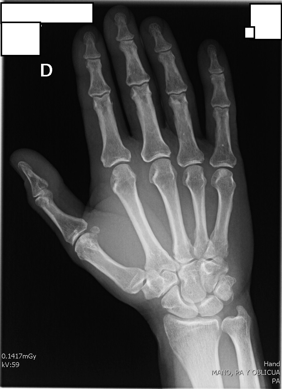
Radiografía PA de Mano - Vista 1
Radiografía PA de Mano - Vista 2
Exposure Factors
45-55
Kilovoltage (kV)
2-4
mAs
100 cm
Distance (SID)
Fine
Focal Spot
No
Grid (Bucky)
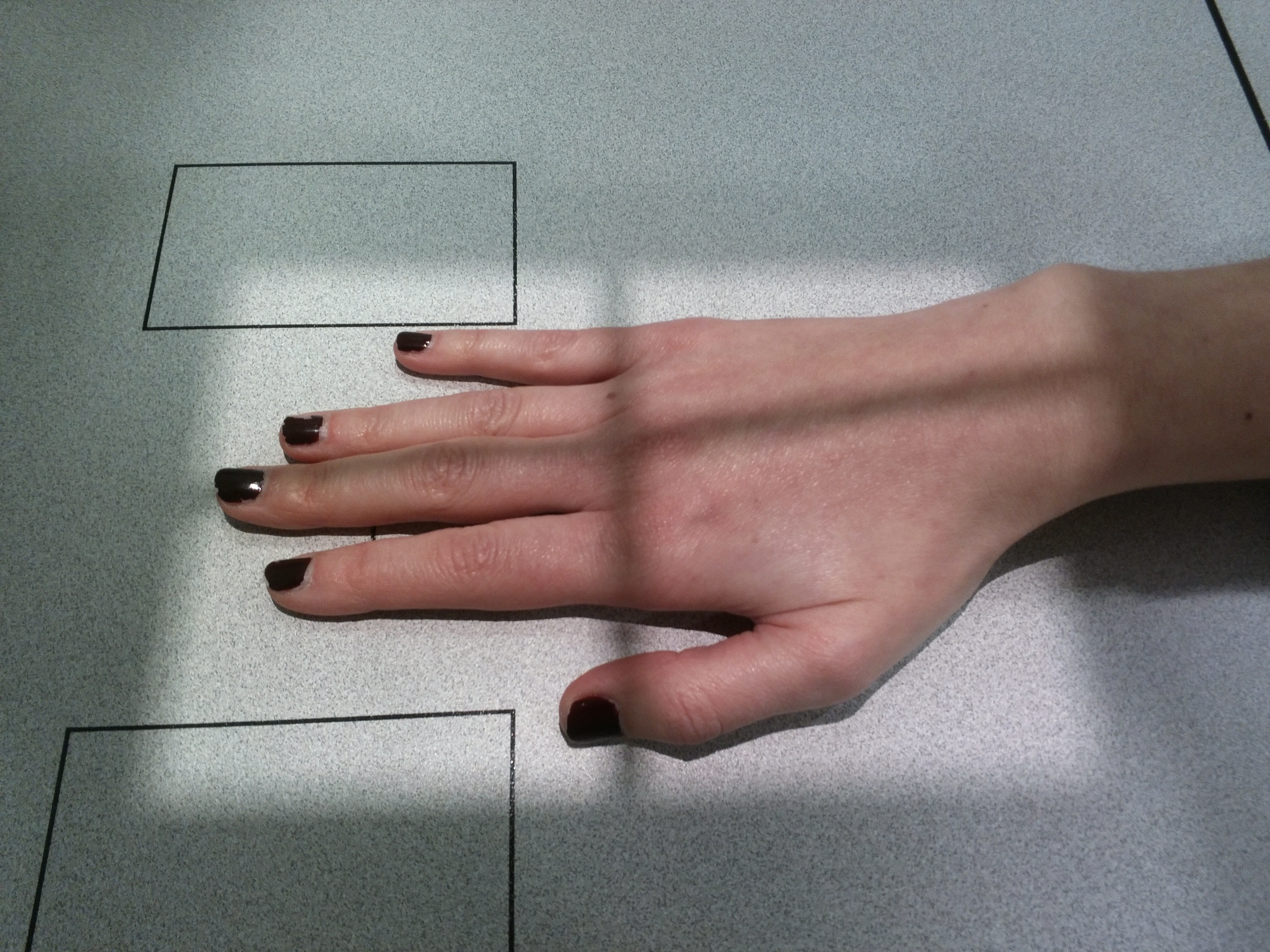
Patient Positioning
Place the hand in prone position on the image receptor.
Spread fingers slightly and ensure the palmar surface is in full contact.
Align the long axis of the hand with the long axis of the IR.
Center the 3rd metacarpophalangeal joint to the IR.
Central Ray
3rd MCP Joint
Location: Perpendicular to the third metacarpophalangeal (MCP) joint.
Visible Anatomy
Phalanges
Distal, middle, and proximal phalanges of all 5 fingers.
Joints
Open IP, MCP, and CMC joint spaces.
Metacarpals
Entire length of the 5 metacarpals.
Standard Radiological Protocol for Hand
Step 1: PA Hand Projection (on top half of the plate)
Step 2: Oblique Hand Projection (on bottom half of the plate)
Step 3: Evaluation of both projections
Step 4: Additional views based on findings (Lateral, Nørgaard)
Step 5: Comparison with contralateral side if necessary